The process of multiplication of a mature mother cell in order to give rise to daughter cells is called cell division. Cell division is central to life of the cell and is essential for the perpetuation of species.
Cell cycle
All those changes which occur during cell growth and cell division are collectively called cell cycle. There are four phases in cell cycle:
- G1 phase (cell growth)
- S phase (DNA replication)
- G2 phase (preparation for division)
- M phase (Cell division) : a) M-Karyokinesis and b) M-Cytokinesis
| Phases | Events within cell |
|---|---|
| G1 (cell growth) | Intensive cellular synthesis, production of structural and functional proteins, cell growth, synthesis of RNA, lipids, carbohydrates. |
| S (DNA replication) | DNA replication, synthesis of histone protein, DNA packaging. |
| G2 (preparation for division) | Synthesis of spindle proteins, ATP molecules, 3 types of RNA (m-RNA, t-RNA, r-RNA) synthesis by DNA, duplication of mitochondria, plastids, centrioles. |
| M- Karyokinesis | Nuclear division in four phases: PMAT (P: prophase, M: metaphase, A: anaphase, T: telophase) |
| M-Cytokinesis | Equal distribution of cell organelles and cytoplasm into each daughter cell. |
Along with these phases, there are also other two stages i.e., checkpoints and Go phase.
Checkpoints (AKA go and stop signals) are where the cell assesses whether conditions are favorable for cell division. There are three checkpoints:
- G1-S transition: DNA damage checkpoint, block the further process if genome is damaged.
- G2-M transition: Initiates mitosis, however block the cycle if DNA replication is uncompleted.
- M-phase transition: Anaphase is blocked if chromatids are not properly assembled on mitotic spindle.
Checkpoints are the guardians of genome which are disrupted in cancer. This rapid cell division results in Cancer.
Go Phase is the inactive phase of cell cycle due to unavailability of mitogens and energy rich compounds. Cells are metabolically active and function as reserve cells.
Types of cell division
There are three types of cell division:
- Amitosis
- Mitosis
- Meiosis
1. Amitosis
Amitosis is the method of growth and development of multicellular organisms. It is also called direct cell division. It is the means of asexual reproduction in bacteria, yeast, protozoans, etc. The following events occur during amitosis:
- Elongation of nucleus
- Formation of constriction in middle
- Deepening of constriction and elongation of nucleus divides it into two daughter nuclei
- Simultaneously, cytoplasm division giving two identical halves.

2. Mitosis
The process of somatic cell division resulting in qualitatively as well as quantitively similar daughter cells with the mother cell. As chromosome number in daughter cell is same as that of mother cell, this division is also called equational division. Especially, it occurs in somatic cells (during cell growth) as well as germ cells of the gonads (during multiplicative phase of gametogenesis).
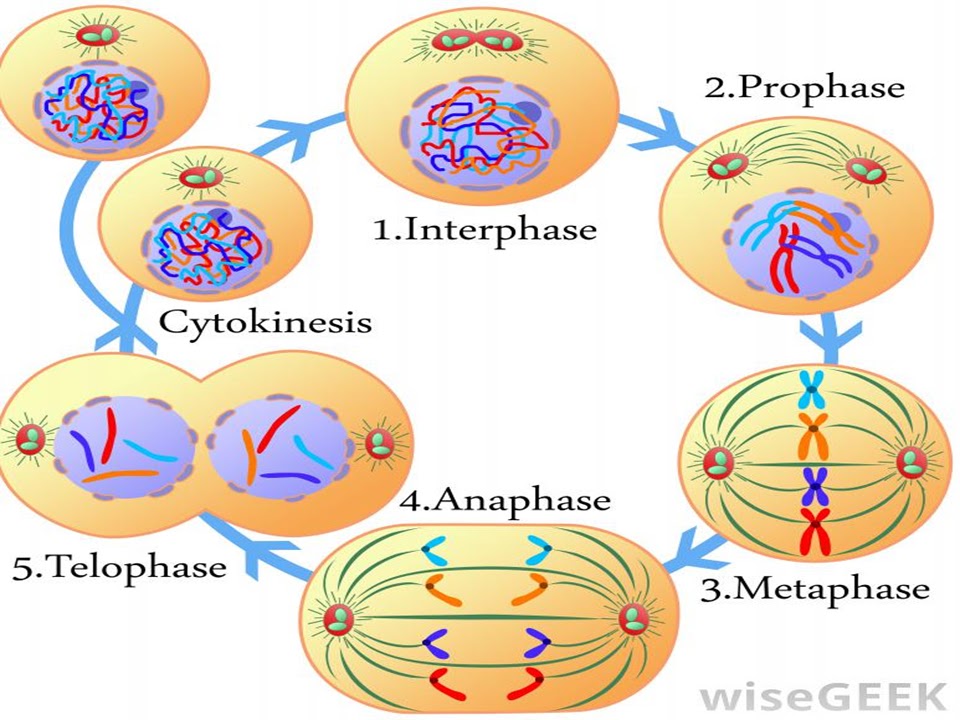
We describe mitosis in following three phases: Interphase, Karyokinesis, Cytokinesis.
A. Interphase
Interphase is the resting stage of cell where cell is metabolically very active. It is the stage between the two successive cell multiplications. The features of the interphase are:
- Cell having large nucleus with intact nuclear membrane and intact nucleolus.
- Diffused, long, coiled and indistinctly visible chromatin fibers.
- DNA duplication into exact copies.
- RNA and protein synthesis.
- Duplication of mitochondria, plastids and centrioles (in animal cell only)
B. Karyokinesis
This process of nuclear division is divided into four phases: Prophase, metaphase, telophase and anaphase.
I. Prophase
- Condensation of the nuclear sap is followed by clearance of nuclear reticulum and nucleoli.
- Distinct chromosomes due to thickening and shortening.
- Double nature of chromosome i.e., chromosome with two sister chromatids and centromere is seen.
- Centriole dividing into two moves towards opposite sides of nucleolus and again divides forming spindle fiber.
- Nuclear membrane and nucleolus begin to disappear by end of this stage.
II. Metaphase
- Complete dissolution of nuclear membrane and nucleolus and simultaneous appearance of spindle fibers.
- Congression; all chromosomes are arranged at the equatorial plane, and centromeres lying at the center of the spindle.
III. Anaphase
- Splitting of centromere so that two sister chromatids of each chromosome has their own centromere forming daughter chromosomes.
- Daughter chromosomes migrate to opposite poles by contraction of spindle fibers and stretching of interzonal fibers.
- Migrating chromosomes have arms directed towards equatorial plane and centromere towards pole.
IV. Telophase
- Each pole receives identical daughter chromosomes.
- Decondensation of chromosomes into chromatin fibers, spindle fibers disappear.
- Reappearance of nucleolus, nuclear membrane around two opposite chromosomal sets.
C. Cytokinesis
Division of extranuclear protoplast is called cytokinesis. It occurs by two ways in different organisms:
I) Cell plate formation : In plant cells, the small granular bodies called phagmoplasts of Golgi complex and ER gather in equatorial region of cell to form a cell plate.
II) Cell Furrowing : In bacteria, fungi and animal cells, a peripheral cleavage furrow appears between two daughter nuclei. Deepening of furrow divides protoplasm into two daughter cells.
Significance of mitosis
- Due to constancy in chromosomal number, mitosis maintains genetic stability or linear heredity.
- It plays vital role in growth and development of zygote into adult.
- It helps in cell repairing, regeneration and also healing of wounds.
- Daughter cells obtained are qualitatively and quantitively identical to mother cells.
- It is the major means of asexual reproduction by fragmentation, budding, stem cutting, etc.
- It also maintains cells size by regulating their division.
3. Meiosis
Meiosis is a special type of cell division of germ cells in sexually reproducing organisms used to produce the gametes, such as sperm or egg cells. It involves two rounds of division that ultimately results in four cells with only one copy of each chromosomes. There are two stages of meiosis: Meiosis-I and Meiosis-II
3.1 Meiosis-I
It is also known as reductional division and divided into two sub-stages: Karyokinesis-I and Cytokinesis-I.
A. Karyokinesis-I
It is also divided into four phases:
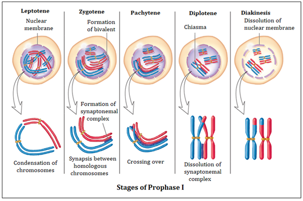
I. Prophase-I
It is the longest and most significant meiotic stage where vital cytogenetical events take place. The five substages of Prophase-I are:
a) Leptotene
- The main event is formation of short, thin, distinct and single stranded chromosomes. Their ends are directed towards the centrosome lying side of nucleus giving bouquet structure.
- Centriole duplicates and migrates to opposite pole and again after reaching the pole, it duplicates and forms diplosomes.
b) Zygotene
- The main event is the synapsis i.e. pairing of homologous chromosomes in a zipper like fashion facilitated by proteinous synaptonemal complex.
- Pairing chromosomes are known as bivalent tetrad chromosomes.
c) Pachytene
- The main event is the crossing over of sister chromatids accompanied by chiasmata formation.
- Initially, the pair of homologous chromosomes spirally twist around each other. And at middle stage, each chromosome divides longitudinally forming 4 sister chromatids in a pair.
- Ligase enzyme holds the broken portions and exchange of chromatin materials occur. This exchange results in the reshuffling, redistribution and mutual exchange of hereditary materials.
d) Diplotene
- The main event is the repulsion of homologous chromosomes. They are attached only at the point of chiasmata.
e) Diakinesis
- The main event is terminalization where chiasma shifts towards the ends of chromosomes and intermediate chiasma diminish.
- Chromatids are still connected by terminal chiasma and they become more condensed and evenly distributed in nucleus.
At late prophase-I , nucleolus detaches from the nucleolar organizer and ultimately disappears. Nuclear membrane disintegrates into vesicles and mitotic spindle fibers begin to form.
II. Metaphase-I
- The main event is the formation of spindle fibers.
- Nucleolus, synaptonemal complex and nuclear membrane disappears.
- Microtubules form spindle fibers attach with the centromere of each tetrad and these homologous pairs lie on equatorial plane.
- Centromere of each chromosome is directed towards opposite pole.
III. Anaphase-I
- The main event is the actual reduction in chromosome number as two sets of haploid chromosomes are formed.
- Each homologous chromosome with two chromatids and undivided centromere separate and move to the opposite poles by repulsive force of bivalents and contraction of spindle fibers.
- Each pole bears chromosomes of either paternal or maternal origin. Due to chiasma formation, two chromatids are genetically unidentical.
IV. Telophase-I
- The main event is the uncoiling and formation of chromatin threads due to decondensation of haploid sets of chromosomes in each pole.
- ER forms the nuclear envelope after disappearance of spindle fibers.
- Nucleolus reappears and thus two haploid nuclei at each pole are formed.
B. Cytokinesis-I
After karyokinesis, cytokinesis occurs and two haploid cells are formed either by cell furrow formation (for animals) or cell plate formation (for plants).
Interphase is absent between meiosis-I and meiosis-II in many cases. Sometimes, it may undergo short interphase only for RNA and protein synthesis but no DNA replication. Hence, S-phase will be absent.
3.2 Meiosis-II
It is also known as equational division and divided into two sub-stages: Karyokinesis-II and Cytokinesis-II.
A. Karyokinesis-II
It has four phases:
I. Prophase-II
- Chromosomes become more thick and short.
- Nucleolus and nuclear membrane disappear again.
- Centriole dividing into two moves towards opposite poles of haploid nuclei and again divides forming spindle fiber.
II. Metaphase-II
- Chromosomes arrange on equatorial plane with spindle fibers attached at centromere.
- New metaphase plate is perpendicular to metaphase plate of meiosis-I.
III. Anaphase-II
- Segregation of sister chromatids after the cleavage of centromere of each chromosome.
- Sister chromatids move to opposite poles by contraction of spindle fibers and now called chromosomes.
IV. Telophase-II
- In each pole, sister chromosomes uncoil into chromatin threads, nuclear membrane and nucleolus reappear and thus haploid four nuclei are formed in total.
B. Cytokinesis-II
Telophase-II is followed by cytokinesis which occurs either by furrowing (for animals) or cell plate formation (for plants). In this way, four haploid cells are formed from single diploid mother cell.

Significance of meiosis
- Meiosis maintains a constant chromosomes number by reducing number of chromosomes of the diploid germ cells into the haploid gametes.
- Crossing over causes genetic variations among species.
- It paves a way for the segregation and independent assortment of genes.
- It is very essential for sexual reproduction.
- It helps in alternation of generation of haploid and diploid generations of plants and animals.
- Failure of leads to formation of polyploid forms.
- It maintains the regularity of reproductive cycle.
Differences between mitosis and meiosis
| Mitosis | Meiosis |
|---|---|
| 1) Mitosis occurs in somatic cells and in germ cells during gametogenesis phase. | 1) Meiosis occurs in reproductive cells of gonads. |
| 2) Completes in one sequence of stages. | 2) Completes in two successive stages of division. |
| 3) Chromosome doubling is followed by single nuclear division. | 3) Chromosome doubling is followed by two nuclear divisions. |
| 4) Daughter cells are genetically identical to mother cell. | 4) Daughter cells are not genetically identical to mother cell. |
| Prophase | Prophase |
| 5) Short prophase with no substages. | 5) Longest prophase in meiosis-I with various substages. |
| 6) No synapsis. | 6) Synapsis in prophase-I. |
| 7) Chromosome duplication takes place in the early prophase. | 7) Chromosome duplication in the late Prophase (Pachytene stage). |
| 8) No crossing over or chiasma formation occurs. | 8) Chiasma formation or crossing over takes place. |
| 9) No exchange of genetic material. | 9) Exchange of genetic material between chromatids of homologous chromosomes. |
| Metaphase | Metaphase |
| 10) Chromosomes appear in the form of dyads or double stranded. | 10) Chromosomes pairs appear in the form of tetrads or four stranded in metaphase-I. |
| 11) As centromere of chromosome divides, its two chromatids become free from each other. | 11) As centromere of homologous chromosomes does not divide, their chromatids do not become free in Metaphase-I. |
| Anaphase | Anaphase |
| 12) Two chromatids of each chromosome separate and migrate to opposite poles of spindle. | 12) Two homologous chromosomes of each tetrad separate and migrate to opposite poles of spindle in Anaphase-I. |
| 13) Daughter chromosomes appear in singlet state. | 13) Daughter chromosomes appear in duplet state in Anaphase-I. However, they split only in Anaphase-II. |
| 14) Long and thin chromosomes. | 14) Short and thick chromosomes. |
| Telophase | Telophase |
| 15) Always present in mitosis. | 15) Sometimes absent in Meiosis-I. |
| 16) The number of chromosome remains same as in mother cell. | 16) Chromosome number becomes half less than the mother cell. |
| Cytokinesis | Cytokinesis |
| 17) Cytokinesis always occurs. | 17) Cytokinesis-I may be absent. |
| Significance | Significance |
| 18) A diploid mother cell produces two diploid cells. | 18) A diploid mother cell produces four haploid cells. |
| 19) Since variations are not produced, it has no role in evolution. | 19) Since crossing over produces some variations, it has a significant role in speciation and evolution. |
References
Leave a Reply