A cell is the basic structural and functional unit of life. In other words, the cell is a unit of biological activity delimited by a selectively permeable membrane and capable of self-reproduction in a medium free of other living systems.
Study of structure, function, molecular organization, growth, reproduction, etc. is called cell biology or cytology.
Discovery of the cell
- The term cell was introduced by an English scientist Robert Hooke in 1665. He observed a thin section of cork using compound microscope. His book Micrographia was published in London in 1665.
- The study of cells began to develop only after the discovery of compound microscope by Jansens in 1590. Because the cells are microscopic structure.
- Anton Von Leeuwenhoek, the famous Dutch microscopist observed chloroplasts in plant cells for the first time. He also studied and observed bacteria and unicellular organisms in 1674.
- In 1831, Robert Brown, an English Botanist described nucleus as a spherical body in plant cells.
- In 1839, Purkinje, the Bohemian physiologist introduced the term protoplasm for the living content of the cell.
- In 1855, Rudolf Virchow stated the fact that all the cells in the body arise from pre-existing cells by cell division.
- Cell wall was investigated extensively in 19th century with the microscope.
- Human eye can see 0.1 micrometer sized object.
Cell Theory
The German botanist, Mathias Jacob Schleiden (1804-1882) and the zoologist Theodar Schwan (1810-1882) first propounded the cell theory. in 1839. It is also called cell doctrine or cell principle. Nigeli, Virchow and Haeckel added and modified the cell theory. The modern cell theory states that-
- Cells are the structural and functional unit of life.
- All living organisms are made up of cells and cell products.
- All cells arise from pre-existing cells of the similar kind.
- Growth of an organism takes place as a result of cell division and cell growth.
- Life passes from one generation to the next in the form of living cells.
- The cells belonging to diverse organisms and different regions of the same organism have a fundamental similarity of structure, chemical composition and metabolism. Simply, all cells are fundamentally similar in chemical compositions and metabolic activities.
- The function of an organism as a whole is the outcome of the activities and interactions of the cells constituting the body of that organism.
- The characteristics and genetic information are stored and expressed inside cells. Hence, cell is the hereditary unit that transfers characters from one generation to another.
- Every cell is made up of a small mass of protoplasm (nucleus, cell organelles, and plasma membrane with or without cell wall).
- Basically, the cells are totipotent unless and until they have become extremely specialized.
- Cells for their survival maintain homeostasis.
- Any organelle in the cell can not survive alone.
Shortcomings of cell theory
The cell theory is more generalized and correct however, it does not apply for all organisms. There are some exceptions:
i) Virus- controversial organism; obligate parasite without cellular organization but has a genetic material (DNA or RNA) surrounded by protein coat.
ii) Bacteria, Cyanobacteria,, Mycoplasma- lack true nucleus, DNA is not enclosed by nuclear membrane but lies directly in the cytoplasm.
iii) Paramecium- Binucleated
iv) Vaucheria(an algae), Mucor, Rhizopus(fungi)- Body made up of coenocytic hyphae(undivided mass of protoplasm in which many nuclei are scattered).
v)Connected cells- Cells are embedded in matrix, a non-living material, nothing is mentioned in theory.
vi) Matured mammalian RBC and sieve tube cells(phloem)- lack nucleus but considered living cells.
The cell as a self contained unit
Cell acts as an autonomous units. The following activities show the independence of the cells:
- Each cell can perform anabolic process and form new structures.
- Every cell is capable of oxidizing food molecules to produce energy and store it in form of ATPs.
- Each cell can reproduce identical daughter cells with same hereditary characters.
- Every cell can respire and exchange gases with surroundings.
- By regulation of its own activities, cell maintains the necessary internal physio-chemical conditions.
- Each cell builds new cell components from its own macromolecules, for the growth and to replace the worn out cells.
- Each cell has its own span.
Unicellular and multicellular organisms
| Unicellular organisms | Multicellular organisms |
| 1. They consist of a single cell to carry out life processes. | 1. They consist of many cells and different cells carry out different functions. |
| 2. Cell size ranges from 11 μm to 1 mm. | 2. Cell size ranges from 5 μm to 100μm. |
| 3. The cell has to be sufficiently large to accommodate all organelles and functions. | 3. Cells are of different sizes and they perform specific functions. |
| 4. They have numerous extensions of cell. | 4. There is the co-ordination of cells to form organ-system. |
| 5. Unicellulars are susceptible to damage even with small injury. | 5. Multicellulars have better capacity and adaptability. |
| 6. They are unable to exhibit wide range of different functions. | 6. They are able to carryout diversified functions. |
| 7. A dead cell for unicellular organism is a dead organism. | 7. Their some of the dead cells also have specific and useful function. |
| 8. Examples: Amoeba, Paramecium, Euglena, etc. | 8. Examples: Rana tigrina, Homo sapiens, Dryopteris, etc. |
Cellular Totipotency
- Totipotency refers to the potential of every living vegetative cell of a plant body to grow into a new plant.
- Haberdtland (1902) first stated that every living plant cell should be able to regenerate a whole plant.
- In 1950, FC Steward with his co-workers gave the concept as well as first experimental evidence of cellular totipotency.
Steward’s experiment to understand the concept of cellular totipotency
- Steward took small pieces from phloem of carrot roots nd put them in a liquid nutrient medium of coconut milk.
- When medium shaken gently, cell clusters spread away from one another.
- Some multiplied and gave rooting clumps.
- They were transferred to another culture tubes having semi-solid medium but same composition.
- It resulted into formation of a new plant which was transferred to a new pot.
- From this experiment, Steward concluded that mature cells, detached from plant body have the capacity to develop into new plants. This is Cellular Totipotency.

Flow of energy through a cell
A constant supply of free energy is required to maintain cellular, tissue, organs, system. Thus, a cell obtains energy from its surroundings either in the form of light(photon) or chemical energy(food molecules) to reduce and avoid entropy. The flow of energy in a cell is as follows:
Light energy (photon) → Chloroplasts → Photosynthesis → Nutrient molecules → Oxidation → ATP synthesis → Food molecules (chemical energy)
Chloroplasts of cells perform photosynthesis in presence of sunlight and the synthesized food molecules are oxidized to form ATP in a living system.
Flow of information through a cell
The flow of information through a cell takes two routes:
i. Flow of intrinsic or genetic information
DNA molecules contain genetic information which control intrinsic activities like metabolism and expression of character by synthesis of protein molecules.
DNA → Transcription → RNA → Translation → Specific proteins
ii. Flow of extrinsic information
Some cells produce hormones which are regulatory substances and combine with cell receptors. Now, their combination controls the metabolic activity of the cell. Additionally, some informational molecules bind with cell membranes molecules and produce cellular effect.
Extrinsic information → Informational molecules → (Membrane receptor →) Cellular receptor → Cellular effects → Proteins
Prokaryotic and eukaryotic cell
| Prokaryotic cell | Eukaryotic cell |
| 1. Incipient nucleus is found. | 1. True and organized nucleus is found. |
| 2. Lack membrane bounded organelles. | 2. Contain membrane bounded organelles. |
| 3. Mitotic apparatus is absent. | 3. Mitotic apparatus is present. |
| 4. Mitotic cell division is absent. | 4. Cell division occurs by mitosis, meiosis and amitosis. |
| 5. DNA lacks histones. | 5. DNA is with histone proteins. |
| 6. Cell wall is made up of murein or peptidoglycan with muramic acid. | 6. Cell wall, if present, is cellulosic. |
| 7. 70s (50s+30s) type ribosome is found. | 7. Both 80s (60s+40s) and 70s ribosomes are present. |
| 8. Respiration occurs by cytoplasmic membranes. | 8. Respiration occurs in the cytoplasm and mitochondria by respiratory enzymes. |
| 9. Cytoplasmic streaming does not occur. | 9. Cytoplasmic streaming occurs. |
| 10. Flagellated prokaryotes do not show 11(9+2) pattern of fibril arrangement. | 10. Flagellated eukaryotes show 11(9+2) pattern. |
| 11. If present, chlorophylls are in lamella not in chloroplasts as thylakoids replace them. | 11. Photosynthetic eukaryotes have membrane closed chloroplasts along with chlorophyll. |
A. Structure of prokaryotic cell

B. Structure of eukaryotic cell

Cell organelles and Cell inclusions
Cell organelles are living sub-cellular structures of the cytoplasm having definite structures and functions. AKA protoplasmic bodies or organoids. They are of two types-
- Organelles related to the chemical work of cytoplasm:
- Mitochondria
- Plastids
- Endoplasmic reticulum
- Golgi complex
- Lysosome
- Ribosomes
- Microsomes
- Microbodies and peroxysomes
- Organelles related to the mechanical work of cytoplasm:
- Microtubules
- Centrosomes
- Flagella and cilia
Cell inclusions are non-living cell bodies of the cytoplasm formed as a result of metabolism in the cell. AKA deutoplasmic or ergastic substances. These can be divided into three categories:
i. Reserve materials ii. Secretory materials iii. Excretory materials.
Parts of eukaryotic cells
Cell wall
- Cell wall is tough, rigid, protective layer found in all plant cells, bacteria, fungi and some protists.
- Absent in all animal cells, some lower plants, male and female gametes and zoospores.
- Thickness varies from 0.1μm to 10μm.
- First discovered by Robert Hooke in 1665 AD.
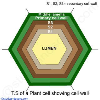
Structure of cell wall
Complex ultrastructure composed of four different layers:
i. Middle lamella
- Thin cementing layer between cell wall of adjacent plant cells formed during cell division.
- Chemically, it is composed of pectates of calcium and magnesium.
- It helps to separate the two adjacent cells.
- The pectin layer of middle lamella is hydrophilic colloidal substance and thus can absorb water.
- The characteristics of softening of ripen fruits is due to partial dissolution of pectic substances and middle lamella.
ii. Primary cell wall
- It is formed by protoplasm on either side of middle lamella.
- Chemically, it is formed of cellulose, hemicellulose and pectin compound.
- It is elastic, thin and permeable.
- Cell walls of most of leaves, fruits, cortex and pith contain only the primary cell wall and primary lamella.
iii. Secondary cell wall
- It is laid down on the primary cell wall.
- It starts developing only after the primary cell wall finished its growth.
- It is composed of three concentric layers of microfibrils laid down one after another.
- The three concentric layers are S1, S2 and S3.
- Chemically, it is composed of lignin in addition to cellulose, hemicellulose and pectin. Some other chemical substances such as cutin, suberin, waxes, mucilage, silica, etc deposited on the wall.
- It is thick, rigid, inelastic and 5μm to 10 μm in thickness.
- Found in certain mature and highly specialized cells such as collenchymatous cells, tracheids, vessels, etc.
iv. Tertiary cell wall
- Layer beneath the secondary cell wall found exceptionally in xylem tracheids of Gymnosperms.
- Chemically, formed of cellulose and xylan and gives additional strength to the cell wall.
- This layer is thin.
v. Plasmodesmata
- These are minute pores present in the primary cell wall.
- also called microscopic channels.
- Help to transport the material from one cell to another.
- Create a pathway for the cell-to-cell communication.
- First reported by Strasburger (1901 AD).
Functions of cell wall
- Mechanical support
- Maintains cell shape.
- Controls cell expansion.
- Controls intercellular transport.
- Protects from infective organisms.
- Suberin and cutin developed in secondary cell wall prevent evaporation.
- Cell wall in root hairs help in absorption.
- Helps from osmotic bursting of the cell.
- Involves in many enzymatic activities.
Plasma membrane
- Fundamental organelle AKA cell membrane/ plasmalemma/ biomembranes.
- Universally present as found in all kinds of cell.
- It is a thin, transparent, electron microscopic, elastic, dynamic, regenerative, living, quasifluidic and selectively permeable membrane.
- Encloses cytosol, nucleus and cell organelles.
- ‘Cell membrane’ given by Nagelli and Cramer in 1855 AD.
- ‘Plasmalemma’ called by Powe in 1931 AD.
- Covers the cell or cell organelles within a cell.
- Chemically, it is lipoproteinous i.e. chiefly composed of lipids (40%), proteins (48-50%) and carbohydrates (2-10%).
Structural models of cell membrane
A. Sandwich model
The model was proposed by Daneili and Davson in 1935. It was based on chemical and physical properties such as permeability to lipid soluble substances. according to this model,
- Lipid bilayer is at the center and protein monolayer and dense is present on either side of the lipid.
- The lipid bilayer is arranged in such a way such that hydrophobic ends directed towards center and hydrophilic ends towards the outer surface. Hence, phospholipid is polar in nature where head is hydrophilic, and tail is hydrophobic.

This model was discarded because:
- It could not explain the physiological properties of the membrane.
- There is not any experimental evidences for the presence of continuous protein layer.
- It could not explain the phenomena like active transport and movement of water-soluble substances.
- It could not explain the functional specificity and variability in the bio membranes.
B. Fluid Mosaic Model
This model was proposed by Singer and Nicholson in 1972. It is based on statement that plasma membrane is formed of protein icebergs floating in the sea of lipid. According to this model,
- Plasma membrane is lipoproteinous and trilaminar.
- Phospholipid bilayer is fluidic in nature because its molecules show two types of movement: a) transition- molecules change position in same layer and b) flip-flop- molecules change position in between two layers.
- Proteins are globular. They are of two types:
- Extrinsic or peripheral proteins- loosely associated and superficially located.
- Intrinsic or integral proteins- tightly associated and penetrate across the bilipid.
- Each phospholipid molecule is a polar molecule and has two specific ends: hydrophilic head of glycerol and hydrophobic tail of two fatty acid chains. So, it is an amphimatic molecule.
- Heads of phospholipid bilayer molecules are directed in opposite directions while the tails of two layers face each other. This arrangement forms a water-resistant barrier.
- Oligosaccharides (carbohydrates molecules) are also associated with the outer surface of plasma-membrane. They either form glycolipids or glycoproteins which help in cellular interaction and in cell-recognition. They also function in blood-grouping, immune response, cancer and rejection of transplanted organs. Cholestor present in some region gives rigidity.
Function of protein in the membrane
- Channel protein facilitate the movement of molecule across the membrane.
- Carrier protein changes its position to help to pass the molecule.
- Receptor protein receive the signals.
- Scaffold protein bind the material and help to maintain the structure.
Functions of cell membrane
- Forms a compartment by separating all the components like cell organelles, nucleus, cytoplasm, etc and work as a unit.
- Protects protoplasm from pathogens, chemicals, etc and also physically.
- The transportation of material through the membrane can be categorized below in 3 ways:
- Passive transport
- Diffusion of molecules: Diffusion is a movement of ions or molecules from a region of higher concentration to a region of lower concentration due to tendency of their molecules to spread uniformly by their random movements. Diffusion helps in exchange of gases in cellular respiration. CO2 and O2 can easily pass through the membrane by diffusion.
- Osmosis: It is process by which solvent (water) migrate across a selectively permeable membrane from a region of higher water potential to region of lower water potential. Cell membrane is semi-permeable where it allows solvent to move in (endosmosis) and move out (exosmosis) without the expenditure of energy.
- Active Transport
- Solute transport: Energy dependent transport (i.e., requires ATP) of solute across a semi-permeable membrane against the concentration gradient. Solute transport can transport the essential solutes such as sugar, amino acids, ions, etc from region of lower concentration to higher one with energy expenditure.
- Bulk Transport
- Endocytosis: It is the process of active intake of large sized or liquid molecules by the membrane inside cell. It is of two types:
- Phagocytosis: Process of intaking large sized particles. (Cell Eating)
- Pinocytosis: Process of intaking large sized liquid nutrients. (Cell Drinking)
- Exocytosis (emeiocytosis/ ephagy/ cell-vomiting): Process of expelling large sized particles outside cell through cell membrane. Those materials are accumulated in exocytic vesicles (membrane bound) which move outward, fuse with plasma membrane and the contents are discharged out.
- Endocytosis: It is the process of active intake of large sized or liquid molecules by the membrane inside cell. It is of two types:
- Metabolism: Various enzymes are present in cell membrane like Lactase, Maltase, etc due to which it is the site of metabolic activities.
- Locomotion: Cell membrane helps in cellular locomotion in two ways which mainly involve the extension and folding of plasma-membrane.
- Pseudopodial movement: Found in amoeba, WBC of blood, macrophages (phagocytes), etc. via finger-like projections.
- Undulatory movement: Found in some mammalian cells such as fibrioblast. Wave-like movement.
- Passive transport
Cell Coat
- The cell coat in animals is also called glycocalyx (glyco=sugar and calyx= outer covering) as it is made of sugars.
- In addition to polysaccharides, many oligosaccharides, glycolipids and glycoproteins can be seen whose composition may vary from cell to cell.
- Cell coat is present on special types of cells and not on others make cell affected by virus, bacteria, hormones and drugs
- Example: most prokaryotes produce polysaccharides that cover their surfaces forming cell coat. These cell coat can also be called capsule, mucilaginous envelope or adhesive coat.
Functions of cell coat
- Mechanical support to the cell.
- Maintains cellular shape.
- Protects the plasma membrane.
- Helps in cell aggregation and tissue formation.
- Involved in histocompatibility.
- Builds immunity against foreign cells making cell sensitive.
- Helps cell-recognition.
- Helps blood-grouping, cancer and rejection of transplanted organs.
Protoplasm (=Bioplasm)
- Fundamental substance of all organisms and considered a living substance.
- A suspension substance that makes up the physical basis of all living things aka building material.
- Term protoplasm was coined by Johannes Purkinje
- Defined as “the physical basis of life” by Huxley in 1868.
- Divided into nucleoplasm (protoplasm of nucleus) and cytoplasm (exo-nuclear protoplasm).
Functions of protoplasm
- Carries on the process of metabolism (synthesis of protein and production of energy).
- Reception of food and oxygen.
- Processes food and oxygen.
- Eliminates waste products.
- Intercellular movement called cyclosis or streaming movement.
Chemical composition of protoplasm
As it is a very colloidal system of substances, it has following composition:
| S.N. | Elements | Percent amount |
| 1. | Oxygen | 62 |
| 2. | Carbon | 20 |
| 3. | Hydrogen | 10 |
| 4. | Nitrogen | 3 |
| 5. | Calcium | 2.5 |
| 6. | Phosphorus | 1.14 |
| 7. | Chlorine | 0.16 |
| 8. | Sulphur | 0.14 |
| 9. | Potassium | 0.11 |
| 10. | Sodium | 0.10 |
| 11. | Magnesium | 0.07 |
| 12. | Iodine | 0.014 |
| 13. | Iron | 0.010 |
Protoplast
- Term coined by Hanstein in 1880 to refer to the entire cell excluding cell wall.
- Can be generated by stripping the cell wall from plant, bacterial or fungal cells by mechanical, chemical or enzymatic means.
Cytoplasm
- Amorphous, translucent, homogenous and colloidal ground substance between cell membrane and nucleus.
- Medium of chemical reaction.
- Provides suitable platform for the operation of other organelles within the cell.
- A site for functions like cell expansion, growth and replication.
- Comprises cytoplasmic matrix (cytosol) and cytoplasmic structures (cell organelles and cell inclusions). Cytoplasmic inclusions include reserve food material, excretory material, secretory material, etc.
- Differentiated into two parts:
- Cytoplasmic matrix
- Cytoplasmic structures
A. Cytoplasmic matrix (Cytosol/Hyaloplasm)
- Consists of inorganic molecules such as water, salts of Na, K and other metals and organic compounds like carbohydrates, lipids, proteins, nucleoproteins, nucleic acids and variety of enzymes.
- Since water makes up 75-85%, aka crystallo-colloidal system (proposed by Kolliker in 1862).
- Peripheral layer is non-granular, viscous, clear, rigid and known as plasmagel or ectoplasm or cortex. Inner portion is granular, less viscous and known as plasmosol, endoplasm or medulla.
Functions of cytoplasmic matrix
- Source of raw materials for functioning of other organelles.
- Site of catabolic pathways e.g.: glycolysis.
- Site of biosynthesis of organic biomolecules like lipids, nucleotides, proteins, etc.
- Helps material exchange between cell organelles and also with environment.
- Helps material distribution in cell.
Cyclosis is an active mass movement of cytoplasm around the central vacuole in one direction or around small vacuoles in several directions. This movement helps in :
- Movement of cell organelles and inclusion.
- Distribution of materials inside the cell.
- Formation of pseudopodia in Amoeba and leucocytes.
B. Cytoplasmic structures (=organelles)
Mitochondria
- Tiny membrane bound cellular structures involved in cellular respiration (release of energy from food).
- Discoverer: Kolliker (1880) in voluntary muscles of insects.
- Term given by C. Benda (1898). GK. mito = thread; chondrion = granule
- Cells of Microsterias, Chlamydomonas has 1 mitochondrion, 5000 in muscle of insect, 300/400 in kidney, 1000/1600 in liver cells.
- Rod-shaped/ filamentous/ spherical/ oval/ globular/ cylindrical/ sausage shaped/ spiral/ cup shape and may change depending on physiological conditions of the cell.
- Largest organelle in animal cell, second largest in plant cell and third largest in cell after nucleus and chloroplast. Average size: 2-6 μm.
Ultrastructure
- Surrounded by two membranes: inner and outer membrane.
- Outer membrane is porous, smooth whereas inner membrane has number of deep folds called cristae providing large surface area for cell respiration.
- These membranes are separated by intermembrane space/ outer chamber filled with watery fluid.
- Space enclosed by inner membrane is matrix or inner chamber which contains dense granules of insoluble inorganic salts, ribosomes, DNA, enzymes of Krebs cycle.
- Mitochondria have their own DNA and RNA.

Functions of mitochondria
- Cellular respiration by the process of catabolism and aerobic respiration.
- Site of biosynthesis of amino acids.
- Regulates Ca ion concentration.
- Helps yolk formation.
- Forms mid piece of sperm.
- Site of synthesis of haeme of hemoglobin and myoglobin.
- Site of thermeogenesis.
- Provides important intermediates for synthesis of chlorophyll, cytochrome, steroids, etc.
Why are mitochondria called the powerhouse of the cell?
Because they release energy in the form of ATP during aerobic respiration for the vital activities of the cell. ATP is source of chemical energy. They intake nutrients, break them down and create energy rich molecules.
Plastid
- Double membranous, semi-autonomous structure found in all plant cells, some protists which is primarily responsible for activities related to manufacture and storage of important chemical compounds (starch, fatty acids, cuticle, epicuticular wax, terpenes, etc.)
- All plastids are derived from protoplastids present in meristematic regions of the plant.
- Protoplastid on division gives different forms of plastid with or without coloring pigment. Pigments are color determiners.
- Plastid is introduced by E. Haeckel in 1866.
- Different types of plastids are:
1. Chloroplast
- Green, double membranous, semi-autonomous structure containing chlorophyll pigment.
- Site of photosynthesis.
- First observer: Antony Von Leeuwenhoek (1679)
- Term given by Schimper (1883)
- Number varies from cell to cell. Chlamydomonas=1, Spirogyra=1-16, plant cell=20-40
- Biconvex/ circular/ cup-shaped/ star-shaped/ ribbon-shaped/ spiral/ reticulate/ filamentous/ ovoid/ discoid, etc
- 2-3 μm thick and 5-10 μm wide.
Ultrastructure
The ultrastructure of chloroplast includes:
- Outer membrane: It is semi-porous membrane permeable to small molecules and ions but not to larger proteins.
- Intermembrane space: It is usually a thin intermembrane space about 10-20 nm present between outer and inner membrane.
- Inner membrane: It is lipoproteinous which forms a border to the stroma. It regulates passage of materials in and out of the chloroplast. In addition, it is also the site of synthesis of fatty acids, lipids and carotenoids.
- Matrix/Stroma: It is dense, colorless, and granular ground substance which is chemically composed of proteins (+50%), 70s ribosomes (plastidoribosomes), DNA (0.5%), mRNA and tRNA, water, minerals (Mn2+, Fe2+, Mg2+) and enzymes. It is the site of dark reaction of photosynthesis. It consists of granum containing thylakoids and intergranal lamella. About 2 to 100 thylakoids in each granum and 100 grana in a chloroplast.
- Thylakoids are disc-shaped, membranous, sacs with a lumen made up of lipid and proteins. It provides platform for light reaction of photosynthesis.
- Intergranal lamella are network of membranous tubules interconnecting grana.
- Chlorophyll pigments are present in thylakoid which can absorb photon of light.
Functions of chloroplast
- Photosynthesis
- Oxygen evolution for aerobic respiration.
- Maintains O2 and CO2 balance in biosphere.
- Prevents global warming
- Starch storage in pyrenoids in algal forms.
- In spinach, site of synthesis of fatty acids
- Responsible for natural greenery.
- Glucose storage and production.
Semi-autonomous nature of chloroplasts
Chloroplasts are semi-autonomous cell organelles because they have a complete machinery to synthesize some of the required proteins while for some other protein synthesis, these depend upon nuclear DNA and cytoplasmic ribosomes.
2. Chromoplasts
- Colored plastids containing varieties of pigments other than green.
- Found in the colored parts of plants like flowers, fruits, etc.
- Formed either from chloroplast by replacement of chlorophyll with other pigments or from leucoplasts by development of pigments.
- Basically, it contains carotenoids (carotene and xanthophyll) pigments. It may also contain anthocyanin dissolved in vacuolar sap of flower petals emitting colors.
- Angular (needle shaped) structures bounded by double layered membrane enclosing stroma without lamella and grana.
- Main function is to
- Attract agents of pollination in flowers.
- Attract agents of dispersal in fruits.
- Types on the basis of pigments present:
- Phaeoplasts (phaeo= dark or brown): Found in diatoms, dinoflagellates, brown algae. Contains brown pigments: Xanthophyll and Fucoxanthin.
- Rhodoplasts: Found in red algae and contains red pigments: R-phucoxanthin.
- Chromatophores of BGA: Found in BGA and contains C-phycocyanin, C-phycoerythrin and Chl-a.
- Chromatophores of photosynthetic bacteria: Present in purple photosynthetic bacteria and contains purple red carotenoid pigments.
3. Leucoplasts
- Leukos- white , plastos- formed
- Colorless, pigment deficient plastids found in storage organs of plant cell unexposed to light. (mainly roots and stems)
- It is like a zip-lock baggie.
- Shape may be rod-like, spherical or oval.
- Double membranous with granular matrix having few lamellae and smaller than chloroplasts.
- They can sometime differentiate into more specialized plastids. Example: transform into chloroplast by developing thylakoids.
- Depending on nature of food stored, its types are:
- Amyloplasts: For starch storage, synthesis and gravity detection. They are found in potato tubers, grains of wheat and rice.
- Elaeoplasts: For lipid (fat or oil) storage. They are found in seeds of mustard, castor, sunflower, cotton, ground nut, etc.
- Proteinoplasts: Protein storing and modifying leucoplasts. They are found in seeds and contain few thylakoids.
- Tannosomes: For synthesizing and producing tannis and polyphenols.
Functions of leucoplasts
- Site of storage of reserve food materials such as carbohydrates, lipids, proteins and oils.
- Site of biosynthesis of fatty acids and amino acids.
Endoplasmic Reticulum
- A well-developed electron microscopic network of interconnected cisternae, vesicles and tubules present throughout the cytoplasm, esp. in endoplasm.
- A membranous network which gives mechanical support to cytoplasmic matrix.
- Garnier in 1897 first observed, Claude and Fullam in 1945 gave ultrastructure and Keith Porter in 1953 gave the name.
- Present in all eukaryotes except matured mammalian RBCs, eggs and embryonic cells. Found more in metabolically active cells such as liver cells, pancreatic cells.
Ultrastructure
Formed by 3 types of elements:
- Cisternae:
- Long, membrane bound, flattened, sac like unbranched structures near the nucleus.
- 40-50 nm in diameter.
- They lie parallel to each other and are interconnected
- Outer surface may bear ribosome due to glycoprotein, ribophorin and fluid filled sac.
- mostly present in cells of pancreas, notochord, brain, etc.
- Vesicles:
- Rounded or oval membrane bound structure found scattered in cytoplasm.
- 25-500 nm in diameter.
- Abundant in pancreatic cells.
- Tubules
- Tubular, irregular and branched structures forming reticulate (net) system along with cisternae and vesicles.
- 50-100 nm in diameter without ribosomes.
- Mainly present near cell membrane and more in lipid forming cells.
Functions
- Makes a network and gives mechanical support to the cytoplasmic matrix.
- Contains enzymes for synthetic and metabolic activities.
- Transportation of the materials: RER -> SER -> GB -> Lysosome
- Increased surface for various enzymatic reaction.
- Detoxification of toxic chemicals by making them more hydrolytic.
- Formation of new nuclear membrane during cell division.
- Storage of synthetic product like glycogen.
- Transfer of information from nucleus to other cell organelles.
- Formation of primary lysosome with hydrolytic enzymes.
- Formation of cell plate during cytokinesis in plant cells.
Differences between rough ER and smooth ER
| S.N. | Rough ER | S.N. | Smooth ER |
| 1. | Ribosomes are present. | 1. | Ribosomes are absent. |
| 2. | Mainly present near the nucleus. | 2. | Mainly present near cell membrane. |
| 3. | Mainly constitute of cisternae. | 3. | Mainly constitute of tubules. |
| 4. | Mainly found in protein forming cells like pancreatic, goblet, plasma cells, Nissl’s granules of nerve cells, etc. | 4. | Mainly found in lipid forming cells like adipocytes, interstitial cells, adrenal cortical, muscle cells, etc. |
| 5. | Formed from nuclear membrane. | 5. | Formed from RER by loss of ribosomes. |
| 6. | Transports and synthesizes protein. It packages polypeptides into proteins and helps in protein transportation. | 6. | Lipids synthesis and storage, carbohydrates metabolism, detoxification of drugs, synthesis of ascorbic acid, steroid hormones, glycogenolysis and phospholipid synthesis. |
Golgi Body/ Golgi Apparatus/ Dictyosomes/ Lipochondrion/ Idiosome/ Dalton Complex
- Part of the endomembrane system in cytoplasm. It packages proteins into membrane-bound vesicles inside the cell before vesicles are sent to their destination.
- Discovered by Camillo Golgi (1898) in nerve muscles of owl.
- Present in all eukaryotic cell except mature sieve elements of plants, mature sperm cell, mature RBC, antherozoids of bryophytes and pteridophytes, etc.
- Shape depends on the functional state of cell. Thus called pleomorphic organelle.
- Size and morphology vary tremendously between different cell types and species. Cisternal membranes and associated vesicles can vary from 0.5 to 2 μm.in diameter.
- Formed from plasmalemma, nuclear envelope and ER.
- Plant Golgi apparatus is composed of many small stacks of cisternae, sometimes known as dictyosomes. Number of stacks and their distribution within the cell is dependent on cell type.
- Maize root cap cells, which are actively secreting large amount of mucopolysaccharides, contains between 300 and 600 Golgi stacks per cell. But there are 25 stacks present in meristematic cells of many plants.
Ultrastructure
- Golgi bodies consist of network of membranes called as cisternae, network of interconnecting tubules, vesicles and vacuoles at peripheral regions (cis and trans faces).
- Cisternae or flattened sacs:
- Elongated, double layered, flat and curved parallel sacs with swollen ends piled one upon other to form stacks.
- Swollen ends are golgian vacuoles.
- Size is about 180-230 Å.
- 3-12 in animal and 10-12 in plant cells.
- Vacuoles:
- Spherical lying towards the maturing face of cisternae.
- Modified and expanded cisternae in which two membranes become widely separated and vacuolar space enlarged.
- Vesicles:
- Intimately attached with the cisternae on its convex surface (forming face).
- Two types:
- Smooth vesicles
- Coated vesicles.
- Cisternae or flattened sacs:
- Golgi body is polar in nature. Its concave side is directed towards cell membrane and called maturation face (trans-face), convex side directed towards nucleus and called forming face (cis-face).
Functions
- Transports, modifies and packages protein and lipids.
- Processes materials to be removed from the cell.
- Makes and secretes mucus.
- Packages products into vesicles for transport.
- Changes, supports and packages the proteins to be stored in cell or secreted out of cell. Hence, it equates to the Post Office or Fed Ex or UPS.
- Helps biosynthesis of glycoproteins.
- Participates in transformation of membranes and recycling of plasma membrane.
- Acrosome of maturing sperm is formed by Golgi body.
- Helps formation of primary lysosome.
- Helps synthesis of pectic substances in cell wall.
- Helps in cell secretion.
- Takes part in cell plate forming during cytokinesis in plant cells.
- Helps formation of hormones in endocrine cells.
Lysosomes
- Lipoprotein membrane bound, sac-like organelles containing digestive enzymes. Consists of 40 types of digestive/ hydrolytic enzymes.
- lysis=digestive ; coma=body.
- Found in eukaryotic cells primarily animal cells and plant cells.
- Discovered by De Duve (1955) who designated them as suicide bags.
- Their primary function is extracellular and intracellular digestion at acidic pH.
- Spherical/ bubble shaped
- Average size is 0.5 μm ranging from 0.2-0.8 μm.
- They breakdown excess of worn-out cell parts. They may be used to destroy invading viruses and bacteria.
- If a cell is damaged before/ beyond repair, lysosomes can help it to self-destruct in a process called programmed death or apoptosis.
- They are known as suicidal bags of cell because it contains digestive enzymes, if something burst, the lysosomes release digestive enzymes which digest all the cells. This leads to death of cells.
- Synthesized in the ER and then transported to Golgi apparatus.
- Has three main functions: the breakdown of unwanted macromolecules, cell membrane repairment, response against foreign substances such as bacteria, viruses and other antigens.
- Lysosomal enzymes are divided into following categories:
- Nucleases: which include ribonuclease and deoxyribonuclease., breakdown polynucleotides into nitrogen bases, phophates and pentose sugars. E.g.: DNase and RNase.
- Phosphatases: Acid phosphate, breakdown phosphate esters to monophosphates. E.g.: ATPase.
- Esterases (lipases): Breakdown lipids into fragments. E.g.: phospholipase, acid lipase.
- Proteases: breakdown protein into their constituent amino acids. E.g.: cathepsin and collagenase.
- Glycosidases: Breakdown into polysaccharides to monosaccharides. E.g.: β-galactosidase, β-glucuronidase, α-mannosidase.
- Sulphatases: Act on sulphatesters, breaking down into fragments. E.g.: sulphate-linked organic compounds.
Types of lysosomes
Lysosomes show polymorphism and can be divided into four types:
- Primary lysosome:
- Storage granules
- Buds from trans face of trans face of Golgi complex or granular ER.
- exogenous materials, deteriorating organelles
- have digestive enzymes in inactive form.
- Secondary lysosomes:
- Heterophagosomes or digestive vacuoles
- Usually look more electron dense
- Contain digestive enzymes and indigested food.
- Residual bodies/ Tertiary lysosomes:
- Telolysosomes
- Lysosomes with only undigested food.
- Thrown out of the cell by exo-cytosis.
- Autophagosomes or autolysosomes:
- Autophagic vacuoles or cytolysosomes
- Formed during the deficiency of foreign food or starvation of the organisms.
- Formed when cell feed on its organelles such as mitochondria and ER by the process of autophagy.
Functions
- Act as cells’ garbage disposal system since they remove dead cells.
- Used for digestion of macromolecules from phagocytosis (ingestion of other dying cells or larger extracellular material, like foreign invading microbes).
- Site for intercellular digestion.
- Play role in endocytosis (where receptor proteins are recycled from the cell surface)
- Help autophagy (auto digestion) wherein old or unneeded organelles or proteins that have invaded the cytoplasm are digested by lysosomal enzymes in autophagosomes.
- Metamorphosis.
- Fertilization.
- Osteogenesis
- Autolysis wherein self-destruction of cell occurs by the release of contents of lysosomes digesting various organelles.
- Heterophagy wherein foreign food/ germ is taken in food vacuole by endocytosis and digested in secondary lysosome.
- Autolysis of cartilage and bone tissue.
Ribosomes
- Dense, granular cellular particle made of RNA and protein that serves as the site for protein synthesis in the cell.
- Smallest known ribonucleic protein particles and universally present for protein synthesis.
- Composed of ribosomal RNA and protein.
- The ribosome reads the sequence of mRNA and using genetic code, translates the sequence of RNA bases into a sequence of amino acids.
- Serve as workbenches, with mRNA acting as the blueprint in the process of protein synthesis.
- Term ribosomes was given by G. Palad (1953)
- Oblate spheroid in shape. One ribosomal unit being larger is dome-shaped and another unit being smaller is cap-like shape.
- 70s ribosome is 200-290 Å * 170-210 Å in size whereas 80s ribosome is 300-340 Å * 200-240 Å in size.
- Number of ribosomes is more in plasma cells, liver cells, meristematic cells, cancer cells, endocrine cells, etc.
- Found in both prokaryotic and eukaryotic cells except mature sperm and RBCs. Eukaryotes have ribosomes in two forms: free (cytoplasmic ribosome) form and bound (on RER and outer nuclear membrane) form.
Types of ribosomes
On the rate ribosomes sediment, they are classified in two types. The heavier the structure, more is its sedimentation coefficient. Some notable differences between two ribosomes are:
| S.N. | 70s | S.N. | 80s |
| 1. | Mostly present in prokaryotic cells, autonomous organelles like mitochondria, plastids, etc. | 1. | Present in eukaryotic cells. |
| 2. | Synthesized in cytoplasm. | 2. | Synthesized in nucleus. |
| 3. | Smaller compared to 80s (about 200 Å in diameter). | 3. | Larger compared to 70s (about 250-300 Å in diameter). |
| 4. | Mostly free floating type. | 4. | May be free floating or membrane bound type. |
| 5. | Has a sedimentation coefficient of 70s. | 5. | Has a sedimentation coefficient of 80s. |
| 6. | Sub-units are 50s (23s+5s+ about 34 proteins) and 30s (16s+ about 21 proteins). Total RNAs=03. | 6. | Sub-units are 60s (28s+5.8s+5s+ about 49 proteins) and 40s (18s+ about 33 proteins). Total RNAs=04. |
| 7. | Protein synthesis of 70s ribosomes is inhibited by cycloheximide. | 7. | Protein synthesis of 80s ribosomes is inhibited by chloramphenicol. |
Functions of ribosomes
- Considered to be protein-factories or engines of the cell because they are the sites of protein synthesis where RNA is translated into protein.
- Co-ordinates and brings all the components of protein synthesis together.
- Carries tRNA from nucleus to the site of protein synthesis. tRNA provides amino acid and anti-codon site.
- Have protective function.
- Free ribosomes produce enzymes for intracellular use.
- Bound ribosomes on RER synthesize enzymes for extracellular use.
- Store proteins temporarily and also rRNAs for protein synthesis.
- Proteins control either cellular structure or act as enzymes and control cellular function. Enzymes also protect mRNA strand.
Nucleus
- Double membranous, most prominent organelle that controls and regulates the activities of the cell and carries the genes, structures that contain the hereditary information.
- It is also called control center of cell (brain of cell) as it contains the genetic information (material) which determines the entire structure and function of the cell.
- It accounts for about 10% of the cell’s volume.
- Either centrally, eccentrically or basally located.
- Shape of nucleus may be spherical, oval, flat, horseshoe, kidney shaped, segmented or lobulated.
- Usually one in eukaryotes, two in liver cells and more than 2 in osteoclasts, skeletal muscle cell.
- Found in all eukaryotes except matured mammalian RBCs, sieve tube cells of phloem, tracheids and vessels of xylem.
- Robert Brown (1831) discovered nucleus.
- Term Nucleolus- Bowman (1840)
- Term Chromatin- W. Flemming (1879)
- Term cytoplasm and nucleoplasm- Strasburger (1882)
Ultrastructure
Nucleus is composed of following four components:
- Nuclear membrane (Karyotheca)
- Thin, transparent membrane, lipoproteinous and trilaminar which separates nucleus from rest of the cell or cytoplasm.
- Discoverer: Erclab (1845)
- It is composed of two outer and inner membranes separated by perinuclear space filled with fluid.
- Perforated by pores which allows for the passage of molecules in and out of the nucleus.
- Outer membrane is continuous with ER and studded with ribosomes.
- Regulates nucleo-cytoplasmic interactions,
- Allows passage of molecules i.e., pinocytosis and phagocytosis.
- Allows passage of ribosomal subunits; mRNA, tRNA and proteins.
- Maintains shape of the nucleus.
- Nuclear sap (Nucleoplasm or Karyolymph)
- A transparent, colloidal, semi-fluid gel like and slightly acidophilic ground substance or matrix inside the nuclear membrane.
- Chemically, made of water, enzyme, lipids, proteins, sugars, minerals (Mn2+, Mg2+), ribosomes, mRNA, tRNA, etc.
- Helps in maintaining shape of nucleus and acts as nuclear skeleton.
- Site of enzyme activities.
- Helps in formation of spindle proteins for cell division.
- Supports nucleoli and chromatin threads.
- Contains 2 types of proteins:
- Basic proteins: They take basic stain. E.g.: Nucleoprotamines, Nucleohistamines.
- Acidic proteins: They are non-histone proteins in nucleoplasm or chromatin. E.g.: Phosphoproteins.
- Chromatin Fibers (Nuclear reticulum)
- Numerous, fine, thread-like, coiled and elongated structures intermingled with one another forming a network.
- Beaded structure of chromatin threads is known as chromomere due to presence of DNA and proteins.
- While cell division, chromatin fibers through spiralization and dehydration condense into number of rods, chromosomes (condensed chromatin gives chromosomes).
- Chromosomes
- They are filamentous bodies present in nucleus which carry the genes (hereditary units) and are visible during cell division.
- They are composed of chromonema, centromeres, secondary constrictions, nucleolar organizers, telomeres and satellites.
- The sub-unit of a chromatid is chromonema. Generally, each chromatid has two chromonemata.
- The localized constricted region of the chromosomes with which spindle fibers attached is centromere or kinetochore or primary constriction. It is functional during cell division. Types of chromosomes on the basis of centromere are: metacentric, sub-metacentric, acrocentric and telocentric.
- The two ends of chromosome are telomeres. They are highly stable and do not unite with other telomeres.
- In addition to primary constriction, the arms of the chromosomes may show one or more secondary constriction II. They represent the sites of breakage and subsequent fusion. The chromosomal region between telomere and secondary constriction is satellite or trapant. Chromosome bearing satellite is satellite chromosome (SAT-chromosome). It helps in identification of chromosomes.
- Constriction near the one end of chromosome is nucleolar organizer because it is necessary for nucleolus formation.
- Functions of chromatin fibers:
- Contain DNA which acts as genetic material.
- Control the synthesis of enzymatic proteins.
- Changes in DNA produce variations.
- 2 types of chromatin:
- Nucleolus:
- A large, spherical, acidophilic, dark and dense granule composed of protein and RNA and generally associated with nucleolar organizer region of nucleolar chromosome.
- Darkly stained granular naked organelle without limiting membrane which consists of granular, fibrillar and amorphous region.
- Helps in spindle fiber formation during cell division.
- Combines the rRNAs and ribosomal proteins to form ribosomal subunits.
| S.N. | Euchromatin | S.N. | Heterochromatin |
| 1. | DNA is loosely packed. | 1. | DNA is tightly packed. |
| 2. | Transcriptionally active. | 2. | Transcriptionally inactive. |
| 3. | Actively present in prokaryotic and eukaryotic genome. | 3. | Only present in eukaryotic genome. |
| 4. | Genetically active. | 4. | Genetically inactive. |
| 5. | Present at inner side of nucleus. | 5. | Present at nucleus periphery. |
| 6. | Stained lighter. | 6. | Stained darker. |
| 7. | Early replicative. | 7. | Late replicative. |
| 8. | Aren’t sticky. | 8. | Are usually sticky. |
| 9. | Allow gene to form protein. | 9. | Regulates genetic integrity, and control gene expression. |
| 10. | Low genetic density. | 10. | High genetic density. |
| 11. | Consist of 2-3 % of genome. | 11. | Consist of 97-98 % genome. |
| 12. | Relatively large amount of DNA. | 12. | Smaller amount of DNA and larger amount of RNA. |
- 4. Nucleolus:
- A large, spherical, acidophilic, dark and dense granule composed of protein and RNA and generally associated with nucleolar organizer region of nucleolar chromosome.
- Darkly stained granular naked organelle without limiting membrane which consists of granular, fibrillar and amorphous region.
- Helps in spindle fiber formation during cell division.
- Combines the rRNAs and ribosomal proteins to form ribosomal subunits.
Functions of nucleus
- Stores genetic information (DNA and RNA) for reproduction, development and behaviour.
- Controls all cellular activities of the cell.
- Controls the synthesis of structural proteins.
- Controls cellular functions by controlling synthesis of enzymatic proteins.
- Houses nucleolus which is the site of production of ribosomes.
- Induces genetic variations which helps in organic evolution.
- Controls cellular differentiation.
- Selective transportation of regulatory factors and energy molecules through nuclear pore.
- DNA replication occurs in the nucleus which is then passed to daughter cells.
Cilia and Flagella
- L. cili- eye lash and flagellum- whip
- Cilia and flagella are fine hair or filamentous protoplasmic structure usually found outside the cell surface but arise internally from the basal body of cytoplasm.
- They are found in both plant and animal cells.
- Cilia are found in ciliated protozoans like Paramecium, body structure such as wind-pipe, kidney, nephrons, etc.
- Flagella are found in flagellated protozoans like Euglena, sperm cells of bryophytes and pteridophytes, zoospores and gametes of several algae and fungi.
Structure
Both cilia and flagella have four parts-
- Basal body:
- Basal part which is embedded in outer part of cytoplasm below the plasma membrane. It is derived from centriole.
- Rootlets:
- Fibrous structures developed from basal body and help to support basal body.
- Basal plate:
- Lies above the basal body at the level of cell membrane. It has 9 (doublet) + 2 (singlet) arrangement of microtubules.
- Shaft:
- Hair-like exposed part of cilia and flagella. It has axoneme surrounded by plasma membrane (sheath).

Functions
- Provide locomotion to the cell or organism.
- Maintain a nutritive-cum-respiratory water current inside the body of Paramecium and sponges.
- Help in elimination of olid particles from repiratory tract.
- Cilia of kidney- nephrons move nephric filtrate.
- Eggs of amphibians and mammals are driven out from the oviduct by cilia.
- Cilia in larval forms help their dispersal.
| S.N. | Cilia | S.N. | Flagella |
| 1. | Extremely fine hair-like small sized 5-10 μm long. | 1. | Fairly long, whip like large sized, upto 150 μm long. |
| 2. | Numerous and may cover the entire surface of the cell. | 2. | Fewer in number and may also occur singly. |
| 3. | Generally distributed on whole body. | 3. | Generally located at anterior end of body. |
| 4. | Beat in either metachronous or synchronous condition. | 4. | Beat independently. |
| 5. | Sweeping or rowing motion. | 5. | Undulatory motion. |
| 6. | Functions: locomotion, feeding, circulation, etc. | 6. | Function: Only locomotion. |
Vacuoles
- Single membranous, fluid filled sacs.
- First reported by Spallanzani.
- Animal cells have numerous and small vacuoles while plant cells have few and large vacuoles. In plants, vacuole is generally large central, or sap vacuole filled with cell sap.
Structure
A vacuole is composed of two parts:
- Tonoplast:
- Outer, single-layered, lipo-proteinous, selectively permeable, unit membrane of about 40 Å thickness.
- Cell-sap:
- Fluid present inside vacuole, formed of water, minerals, sugars, amino acids, proteins, esters, anthocyanins, organic acids, O2, CO2 and some wastes, secondary products of metabolism.
Vacuoles can modify differently to perform specific functions. E.g.: food vacuoles, contractile vacuoles and gas vacuoles (pseudo vacuoles), etc.
Functions
- Stores water, minerals, compounds like sugars, amino acids, pigments, waste products, plant metabolites (tannin and latex), etc.
- Makes plant cell turgid.
- Helps cell growth and elongation.
- Contractile vacuole is osmoregulatory in function and helps excretion in protozoans.
- Gas vacuoles in cyanobacteria provide buoyancy.
Cell inclusions
- Metabolically inactive non-living substances present in cytoplasm.
- AKA deuteroplasmic bodies, ergastic bodies.
- E.g.: proteins, amino compounds, starch grains, essential oils, latex, mineral crystals, cellulose, insulin, sugars, fats, oils, pigement, enzymes, etc.
- Divided into 3 categories:
- Reserve materials: Metabolic products stored in plant cells from the reserve food of the plant.
- Nitrogenous reserve food materials: They always have some amount of nitrogen and are present in almost every cell. E.g.: proteins, amino compounds.
- Non-nitrogenous reserve food materials: Only contain carbon, hydrogen and oxygen. They are the products of photosynthesis. E.g.: starch, cellulose, sugars, insulin, fat and oils, glycogens, etc.
- Secretory materials: Certain substances formed during plant metabolism and are not concerned with nutrition. They are of following types:
- Plant pigments: Color producing substances of plants. E.g.: chl-a, chl-b, chl-c, etc.
- Enzymes: Nitrogenous secretory products. AKA biocatalysts.
- Nectar: Secreted from nectaries present near or at the base of ovary or stamens of flowers. It helps in pollination.
- Excretory materials: By-products of plant metabolism and are of no use to plants. Examples: alkaloids, organic acids, gums, resins, tannins, essential oils, latex, mineral crystals, etc.
- Reserve materials: Metabolic products stored in plant cells from the reserve food of the plant.
References
Fig.1 : Cellular Autonomy and Totipotency | Class Eleven Biology (askmattrab.com)
Fig.2 : Prokaryotic Cells – Blog (weebly.com)
Fig. 5: sandwich model of plasma membrane – Bing images
Fig. 6: https://bichep.com/chemical-nature-of-protoplam-bichep-com/
Fig.7: https://getrevising.co.uk/revision-cards/cell-ultrastructure-2
Fig.8: https://www.studyandscore.com/studymaterial-detail/chloroplast-distribution-morphology-ultrastructure-and-functions
Fig.9: https://www.thoughtco.com/endoplasmic-reticulum-373365
Fig.10: https://rsscience.com/golgi-apparatus-function-the-post-office-inside-the-cells/
Fig.11: https://kullabs.com/index.php/class-11/biology11/cell-organelles/lysosomes
Fig.12: https://www.quora.com/What-is-the-function-of-the-70S-and-80S-ribosomes
Fig.13: https://funscience.in/nucleus/
Fig.14: https://cloudshareinfo.blogspot.com/2019/08/difference-between-cilia-and-flagella.html
Leave a Reply